Drinking Coffee: Study Finds Both Beneficial and Harmful Short-Term Health Effects

This article is a repost which originally appeared on SciTechDaily
AMERICAN HEART ASSOCIATION - JANUARY 19, 2022
Edited for content and readability - Images sourced from Pexels
Source: https://clinicaltrials.gov/ct2/show/study/NCT03671759

Key Points:

  • A randomized trial to study caffeinated coffee consumption among 100 volunteers for two weeks found both potentially beneficial and harmful short-term health consequences of drinking coffee.
  • When participants were randomly assigned to drink coffee, they were more physically active, yet they also had an increased number of abnormal heartbeats and slept less compared to when they were randomly assigned to avoid all caffeine.

Drinking caffeinated coffee appears to have both beneficial and harmful short-term health effects: increased abnormal heartbeats, increased physical activity, and reduced sleep duration, according to research that was presented at American Heart Association’s Scientific Sessions 2021.

“Coffee is the most commonly consumed beverage in the world, yet its health effects remain uncertain,” said study author Gregory Marcus, M.D., M.A.S., associate chief of cardiology for research and endowed professor of atrial fibrillation research at the University of California, San Francisco. “While the majority of long-term observational studies have suggested multiple potential benefits of drinking coffee, this is the first randomized trial to investigate the real-time, physiologic consequences of coffee consumption.”

Marcus and colleagues enrolled 100 adult volunteers, and they were assigned to wear continuously recording ECG devices (to track heart rhythm), wrist-worn devices to track physical activity and sleep; and continuous glucose monitors to track blood sugar levels for two weeks. The participants were an average age of 38 years, 51% were women and 48% were white. Researchers also obtained DNA saliva samples from the participants to assess genetic variants that may affect caffeine metabolism.

Participants were then randomly assigned to either avoid or consume coffee for no more than two consecutive days each for 14 consecutive days. Coffee and espresso consumption were recorded in real time via a “time stamp button” on the ECG monitor, and researchers tracked trips to coffee shops with geotracking. In addition, participants completed daily questionnaires to detail how much coffee they had consumed every morning.

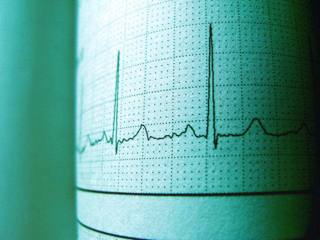
The analysis found that coffee consumption was associated with a 54% increase in premature ventricular contractions, a type of abnormal heartbeat originating in the lower heart chambers reported to feel like a skipped heartbeat. In contrast, drinking more coffee was associated with fewer episodes of supraventricular tachycardia, an abnormally rapid heart rhythm arising from the upper heart chambers.

Consuming coffee was consistently associated with more physical activity as well as less sleep. Specifically:

  • Participants who consumed coffee logged more than 1,000 additional steps per day compared to days when they did not drink coffee.
  • On the days participants drank coffee, they had 36 fewer minutes of sleep per night according to their Fitbit devices.
  • Drinking more than one coffee drink more than doubled the number of irregular heartbeats arising from the heart’s lower chambers.
  • Each additional cup of coffee consumed was associated with nearly 600 more steps per day and 18 fewer minutes of sleep per night.
  • There were no differences in continuously recorded glucose measured when the study participants consumed versus avoided coffee.

These findings were corroborated by analyses of adherence to their randomization assignment and amplified when more versus less coffee was consumed.

“More physical activity, which appears to be prompted by coffee consumption, has numerous health benefits, such as reduced risks of Type 2 diabetes and several cancers, and is associated with greater longevity,” Marcus said. “On the other hand, reduced sleep is associated with a variety of adverse psychiatric, neurologic and cardiovascular outcomes. More frequent abnormal heartbeats from the upper heart chambers influence risk of atrial fibrillation, and more frequent abnormal beats from the lower chambers, or ventricles, increase the risk of heart failure. These results highlight the complex relationship between coffee and health.”

The study participants with genetic variants associated with faster caffeine metabolism exhibited more abnormal heart beats originating in the ventricles, or PVCs, when more caffeinated coffee was consumed. The slower an individual metabolized caffeine based on their genetics, the more sleep they lost when they drank caffeinated coffee.

The investigators also sought to determine if changes in exercise or sleep influenced coffee’s effects on abnormal heart rhythms, and no such association was identified.

Marcus noted that because coffee was randomly assigned to the study participants, cause-and-effect can be inferred. These observations were made during repeated assessments of days when coffee was consumed versus when it was not for each study participant, eliminating concerns regarding differences in individual-level characteristics as an explanation for these results.

Scientists show how AI may spot unseen signs of heart failure

A special artificial intelligence (AI)-based computer algorithm created by Mount Sinai researchers was able to learn how to identify subtle changes in electrocardiograms (also known as ECGs or EKGs) to predict whether a patient was experiencing heart failure.

This article is a repost which originally appeared on Medical Xpress
The Mount Sinai Hospital - October 19, 2021 
Edited for content and readability Images sourced from Pexels 
DOI: https://www.jacc.org/doi/10.1016/j.jcmg.2021.08.004

“We showed that deep-learning algorithms can recognize blood pumping problems on both sides of the heart from ECG waveform data,” said Benjamin S. Glicksberg, Ph.D., Assistant Professor of Genetics and Genomic Sciences, a member of the Hasso Plattner Institute for Digital Health at Mount Sinai, and a senior author of the study published in the Journal of the American College of Cardiology: Cardiovascular Imaging. “Ordinarily, diagnosing these type of heart conditions requires expensive and time-consuming procedures. We hope that this algorithm will enable quicker diagnosis of heart failure.”

The study was led by Akhil Vaid, MD, a postdoctoral scholar who works in both the Glicksberg lab and one led by Girish N. Nadkarni, MD, MPH, CPH, Associate Professor of Medicine at the Icahn School of Medicine at Mount Sinai, Chief of the Division of Data-Driven and Digital Medicine (D3M), and a senior author of the study.

Affecting about 6.2 million Americans, heart failure, or congestive heart failure, occurs when the heart pumps less blood than the body normally needs. For years doctors have relied heavily on an imaging technique called an echocardiogram to assess whether a patient may be experiencing heart failure. While helpful, echocardiograms can be labor-intensive procedures that are only offered at select hospitals.

However, recent breakthroughs in artificial intelligence suggest that electrocardiograms—a widely used electrical recording device—could be a fast and readily available alternative in these cases. For instance, many studies have shown how a “deep-learning” algorithm can detect weakness in the heart’s left ventricle, which pushes freshly oxygenated blood out to the rest of the body. In this study, the researchers described the development of an algorithm that not only assessed the strength of the left ventricle but also the right ventricle, which takes deoxygenated blood streaming in from the body and pumps it to the lungs.

“Although appealing, traditionally it has been challenging for physicians to use ECGs to diagnose heart failure. This is partly because there is no established diagnostic criteria for these assessments and because some changes in ECG readouts are simply too subtle for the human eye to detect,” said Dr. Nadkarni. “This study represents an exciting step forward in finding information hidden within the ECG data which can lead to better screening and treatment paradigms using a relatively simple and widely available test.”

Typically, an electrocardiogram involves a two-step process. Wire leads are taped to different parts of a patient’s chest and within minutes a specially designed, portable machine prints out a series of squiggly lines, or waveforms, representing the heart’s electrical activity. These machines can be found in most hospitals and ambulances throughout the United States and require minimal training to operate.

For this study, the researchers programmed a computer to read patient electrocardiograms along with data extracted from written reports summarizing the results of corresponding echocardiograms taken from the same patients. In this situation, the written reports acted as a standard set of data for the computer to compare with the electrocardiogram data and learn how to spot weaker hearts.

Natural language processing programs helped the computer extract data from the written reports. Meanwhile, special neural networks capable of discovering patterns in images were incorporated to help the algorithm learn to recognize pumping strengths.

“We wanted to push the state of the art by developing AI capable of understanding the entire heart easily and inexpensively,” said Dr. Vaid.

The computer then read more than 700,000 electrocardiograms and echocardiogram reports obtained from 150,000 Mount Sinai Health System patients from 2003 to 2020. Data from four hospitals was used to train the computer, whereas data from a fifth one was used to test how the algorithm would perform in a different experimental setting.

“A potential advantage of this study is that it involved one of the largest collections of ECGs from one of the most diverse patient populations in the world,” said Dr. Nadkarni.

Initial results suggested that the algorithm was effective at predicting which patients would have either healthy or very weak left ventricles. Here strength was defined by left ventricle ejection fraction, an estimate of how much fluid the ventricle pumps out with each beat as observed on echocardiograms. Healthy hearts have an ejection fraction of 50 percent or greater while weak hearts have ones that are equal to or below 40 percent.

The algorithm was 94 percent accurate at predicting which patients had a healthy ejection fraction and 87 percent accurate at predicting those who had an ejection fraction that was below 40 percent.

However the algorithm was not as effective at predicting which patients would have slightly weakened hearts. In this case, the program was 73 percent accurate at predicting the patients who had an ejection fraction that was between 40 and 50 percent.

Further results suggested that the algorithm also learned to detect right valve weaknesses from the electrocardiograms. In this case, weakness was defined by more descriptive terms extracted from the echocardiogram reports. Here the algorithm was 84 percent accurate at predicting which patients had weak right valves.

“Our results suggested that this algorithm may eventually help doctors correctly diagnose failure on either side of the heart,” Dr. Vaid said.

Finally, additional analysis suggested that the algorithm may be effective at detecting heart weakness in all patients, regardless of race and gender.

“Our results suggest that this algorithm could be a useful tool for helping clinical practitioners combat heart failure suffered by a variety of patients,” added Dr. Glicksberg. “We are in the process of carefully designing prospective trials to test out its effectiveness in a more real-world setting.”