
Vitamin D is a fat-soluble essential vitamin that our skin synthesizes when exposed to the sun. It benefits us in many ways, from bone health to mood.
Summary of Vitamin D
Primary Information, Benefits, Effects, and Important Facts
Vitamin D is a fat-soluble nutrient. It is one of the 24 micronutrients critical for human survival. The sun is the major natural source of the nutrient, but vitamin D is also found naturally in fish and eggs. It is also added to dairy products.
Supplemental vitamin D is associated with a wide range of benefits, including increased cognition, immune health, bone health and well-being. Supplementation can also reduce the risks of cancer, heart disease, diabetes and multiple sclerosis. People deficient in vitamin D may also experience increased testosterone levels after supplementation.
The body produces vitamin D from cholesterol, provided there is an adequate amount of UV light from sun exposure. There is only a sufficient amount of UV light coming from the sun when the UV index is 3 or higher, which only occurs year-round near the equator, between the 37th parallels.
Most people are not deficient in vitamin D, but they do not have an optimal level of vitamin D either. Due to the many health benefits of vitamin D, supplementation is encouraged if optimal levels are not present in the body.
Things To Know & Note
Is a Form Of:
Primary Function:
Do Not Confuse With:
Calcitriol or 1, 25-Dihydroxyvitamin D
(Hormonally active yet not directly supplemented form)
Other Functions:
- Antioxidant and Anti-inflammatory
- Allergies and Immunity
- Bone and joint health
- Cognitive Function and Brain Health
- Insulin Sensitivity
- Cardiovascular
- Libido and Sexual Health
Also Known As:
Cholecalciferol (Vitamin D3), Ergocalciferol (Vitamin D2)
Goes Well With:
- Calcium (for bone health)
- Vitamin K (for bone health, and vitamin K
may attenuate the risk for vitamin D overdosing)
- Vitamin D is usually seen as non-stimulatory
- Vitamin D may have enhanced absorption when taken with meals
How to Take Vitamin D
Recommended dosage, active amounts, other details
The recommended daily allowance for Vitamin D is currently set at 400-800IU/day, but this is too low for adults. The safe upper limit in the United States and Canada is 4,000IU/day. Research suggests that the true safe upper limit is 10,000IU/day. For moderate supplementation, a 1,000-2,000IU dose of vitamin D3 is sufficient to meet the needs of most of the population. This is the lowest effective dose range. Higher doses, based on body weight, are in the range of 20-80IU/kg daily.
Vitamin D3 supplementation (cholecalciferol) is recommended over D2 supplementation (ergocalciferol), since D3 is used more effectively in the body.
Vitamin D should be taken daily, with meals or a source of fat, like fish oil.
Frequently Asked Questions about Vitamin D
Q: When should I take Vitamin D?
Q: Can vitamin D-crease pain?
Q: How can you increase testosterone naturally?
Q: Do I need to supplement Vitamin D if I drink fortified milk?
Q: Can vitamin D cure depression?
Human Effect Matrix
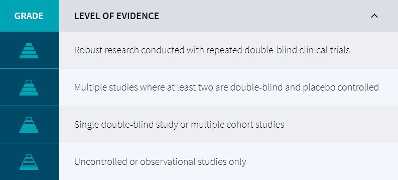
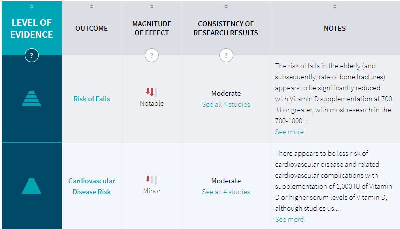
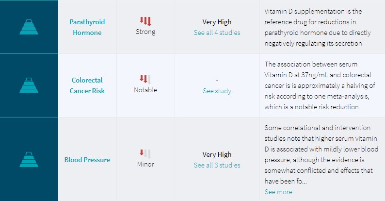
The Human Effect Matrix looks at human studies (it excludes animal and in vitro studies) to tell you what effects vitamin d has on your body, and how strong these effects are.


Studies Excluded from Consideration
- Used injections of the active hormone[1]
Scientific Research on Vitamin D
1. Sources and Structure
1.1 Sources and Intake
Vitamin D is a compound classified as an essential vitamin that derives its name from simply being discovered shortly after Vitamins A, B (prior to the realization that Vitamin ‘B’ was not a single molecule), and Vitamin C.[2] It was initially found to be a component of Cod Liver Oil, and credited as the ‘anti-rachitic’ (against rickets) compound to explain how Cod Liver Oil was effective in treating rickets.[3]
Vitamin D is a term used to refer to a group of related molecules, which collectively increase the body’s pool of 25-hydroxyvitamin D (circulating form of Vitamin D) and subsequently 1,25-dihydroxyvitamin D (the active hormone).
Food sources of vitamin D3 include:
- Milk, being the most common source of vitamin D in the USA[4] and has trended downwards in recent decades[5]
- Cod liver oil at around 2.54-2.78mcg/mL;[6] although labels would be more precise on a product-specific basis as some are lower than this estimate such as 33.5-172.3IU/mL[7][8]
Dairy appears to be the best food source for vitamin D3. Cod liver oil effectiveness varies, depending on the processor and the method of analysis.
Previously, the RDA was set at 400IU in 1997 (International Units, approximately equal to 10mcg Vitamin D3) as this dose is sufficient to reduce the risk of rickets in children.[9] Even currently, an intake of 400IU (despite keeping the mother in a clinically ‘deficient’ state according to current definitions) is sufficient to prevent the occurrence of rickets.[10]
This 400IU target intake, as well as the actual overall intake of Vitamin D3, is commonly seen as deficient in adults[11] as 400IU cannot ideally sustain circulating levels between 50-75nmol/L, which is seen as ideal.[12][13]
1.2 Synthesis from the Sun
Synthesis of vitamin D can occur after contact with the sun, where bodily stores of 7-dehydrocholesterol (a derivative of cholesterol) converts to cholecalciferol (vitamin D3).[14]
In some scenarios, the rate of vitamin D synthesis is reduced; such as:
- Latitudes that are further away from the equator tend to reduce synthesis rates due to less exposure to solar radiation. Several studies note that Northern USA (relative to Southern USA) experience less UVB radiation[15], which appears to be related to cancer risk[16]
- Weather patterns or seasons that reduce solar exposure, such as clouds or darkness[17][18]
- A combination of latitude and season, with the Northern Hemisphere (Boston and Edmonton; latitude 42.2-55°N) failing to produce any vitamin D between October and March[19]
- Areas with higher ozone breakdown (assessed by Dobson units) appear to have higher UVB radiation[20]
- Darker skin has a slower synthesis rate than lighter skin and Black persons are routinely at a higher risk for vitamin D deficiency when compared to lighter skin tones (Asian, Caucasian, Hispanic) when other factors are controlled for.[21][22]
Several factors listed above influence the rates of vitamin D synthesis from the sun. The two most relevant factors are latitude, since being closer to the equator results in more vitamin D synthesis, and skin tone, with black people having a higher risk of vitamin D deficiency. An outright failure to produce any UVB-induced previtamin D has been noted above latitude 42.2°N (Boston) from November to February (4 months), which is prolonged to 6 months above latitude 55°N (Edmonton). The range of 18-32°N still produces vitamin D during the winter.
Despite some sunscreen potentially being related to reduced cancer risk from the sun (Melanoma),[23][24] a topic that is somewhat mixed in results,[25] sunscreens appear to significantly attenuate synthesis of vitamin D by interfering with the topical influences of UVB rays.[26][27] Chronic (not acute) sunscreen usage has been associated with Vitamin D deficiency.[28]
Sunscreen is able to significantly diminish synthesis of vitamin D, and chronic usage may be associated with vitamin D deficiency, if no oral supplementation exists.
1.3 Structure
The standard supplement is vitamin D3, otherwise known as cholecalciferol; vitamin D3 tends to be better absorbed than other forms of vitamin D. In the liver, cholecalciferol is turned into 25-hydroxycholecalciferol via the enzyme cholecalciferol 25-hydroxylase and then sent out to the kidneys to get hydroxylated into 1,25-dihydroxycalciferol. 1,25-dihydrocalciferol is also known as Calcitriol, and is the active hormone that is the result of vitamin D3 ingestion.

1.4 Bioactivation
Vitamin D is known to be a steroid precursor, which implies that it is currently not bioactive but can become active in the body after metabolism. There are different pathways for oral supplementation and biological synthesis originating in the skin.
When supplementation is not relevant, bodily stores of 7-dehydrocholecalciferol must be converted to cholecalciferol (Vitamin D3). This initial metabolite is present in the skin and the metabolism is initiated by light (spectrum 280-320 UVB[29]) which breaks a part of the molecule known as the B-ring. The metabolite, called pre-vitamin D3, then isomerizes to Vitamin D3 and can then be subject to metabolism in the liver.[29][30]
The first stage of bioactivation from the molecule cholecalciferol towards the product 25-hydroxycholecalciferol is mediated by a 25-hydroxylase, both CYP2R1 and CYP27A1 being implicated.[14] This process occurs primarily in the liver and due to the next enzyme (CYP27B1) being primarily expressed in the kidneys a large amount of 25-hydroxycholecalciferol is ejected into serum so it can reach this tissue. Upon being subject to CYP27B1 the product is then converted into 1,25-dihydroxycholecalciferol which is considered the active form of Vitamin D as a hormone.[14]
Vitamin D3 is bioactived into its hormone form in either two stages (if starting from a dietary supplement containing Vitamin D3) or in three stages if starting from skin storages, with the skin mediating the first (nonsupplemental) conversion and the later two metabolic steps being handled in the liver and kidneys respectively
1.5 Variants of Supplementation
Vitamin D itself is divided into two forms, ergocalciferol (vitamin D2), which is mostly derived from plants, and cholecalciferol (vitamin D3), which is the form produced in mammals and fish and thus is a component of cod liver oil supplementation (alongside vitamin A and fish oil fatty acids).[31][32] The only difference in these two molecules is a methyl group, as vitamin D3 is 27 carbons in length, while D2 is 28 carbons.[33]
Both vitamin D2 and D3 are seen as prohormone compounds (acting to increase circulating levels of 25-hydroxyvitamin D)[31] although it appears there is controversy over which form is superior in increasing circulating 25-hydroxyvitamin D, with many sources suggesting that vitamin D3 is more effective as the active hormone is 25-hydroxycholecalciferol rather than ergocalciferol (more closely resembling that of D3 than D2 in structure) and that D2 should not be sold as a supplement.[34]
Due to the differences in molecular weight one IU of vitamin D3 is 25ng in weight, while one IU is 25.78ng in weight (the difference being the aforementioned methyl group) meaning that a dose of 400IU for vitamin D3 (10µg) would be 385IU, and this difference was thought to be significant for the prevention of rickets and food fortification.
Vitamin D2 and D3 are two forms of vitamin D supplementation that are capable of increasing circulating levels of the active hormone. Although D3 is more potent than D2 (based on weight), it is (controversially) thought that standardizing the two to an IU value normalizes the difference.
Some studies, such as 11 weeks of supplementation at the winter at 1,000IU (D2 or D3, with a third group given 500IU of each) either as supplementation[35] or orange juice fortification[36] have noted equivalence between the two forms, and elsewhere supplementation of 1,000IU daily in vitamin D deficient persons has noted a difference in circulating hormone levels but no differences in parathyroid hormone.[37]
In a few cases, supplementation of vitamin D2 have increased levels of the molecule 1,25-dihydroxyergocalciferol yet reduced circulating levels of 1,25-dihydroxycholecalciferol.[38]
Other studies using daily dosing of 1,600IU for a year,[33] 4,000IU over 14 days,[39], and for intermittent doses 50,000IU one monthly for a year[33] or a one time dose[40] as well as acute doses of up to 300,000IU D3[41] have also been noted to be more effective than D2, and according to a meta-analysis the difference in efficacy between D3 and D2 is more notable with bolus supplementation than with daily supplementation.[42]
When comparing D2 against D3 (on an IU basis), there is mixed evidence for both supplements suggesting either bioequivalence (no significant difference) or a superiority of vitamin D3. As there are no studies suggesting that D2 is more efficient, it would be prudent to choose D3 supplementation.
D2 is synthetically produced (for the purpose of supplementation) from irradiation of ergosterol (from mold ergot) whereas D3 is synthesized from 7-dehydrocholesterol.[43]
D2 appears to be less chemically stable than D3 ex vivo (not in an oil base, however)[44][45]leading to some authors suggesting that it may have a poorer shelf-life.[34]
D2 and D3 are synthesized (for supplementation) by different means, and there appear to be differences in their stability, with D3 being more stable than D2, in powder form.
2 Circulating Vitamin D Levels
2.1 Target Levels
Currently, the generally accepted terms to refer to different possible ‘states’ of Vitamin D status are:[46]
- Deficiency (Less than 30nmol/L or 12ng/mL, leading to rickets in children and osteomalacia in adults)
- Insufficieny (between 30-50nmol/L, the range of 12-20ng/mL)
- Adequate (between 50-125mol/L, or 20-50ng/mL)
- High (above 125nmol/L or 50ng/mL)
(where 2.5 nmol/L is approximately equal to 1 ng/mL, and 1 microgram (mcg or µg) of Vitamin D3 is approximately 40IU[47])
The above are generally accepted guidelines for vitamin D, and will be used as reference for this article. ‘Optimal vitamin D levels’ is not a legitimate term to refer to one of these four ranges.
A target of 75nmol/L has been considered to as optimal for bone health in older individuals[48] and bone-related conditions such as dental health or reducing the risk of falls and fractures in the elderly.[49] This also appears to be a target for colorectal cancer prevention.[49]
Even in studies that recommend higher oral intakes (5,000IU), the end goal still appears to be around 75-80nmol/L.[11]
2.2 Deficiency (Predictors)
Using other cut-offs, in 2010 29% of the American population was below 50nmol/L (clinical insufficiency) and 3% below 20nmol/L (clinical deficiency).[51] These levels vary by season, and using 50nmol/L as a cut-off again, 11% of people are below this line at the end of summer (testing area of Boston, latitude 42°N), while at the end of winter this number increases to 30%.[52] In a slightly more northern region on the other side of the globe (Britain, latitude 53.1°N) rates of deficiency still increase. When assessing the serum levels of 25, 50, and 75nmol/L the percentage of the population having less than these values increases from 3.2%, 15.4%, and 60.9% at the end of summer to 15.5%, 46.6%, and 87.1% at the end of winter.[53] Estonia (59°N) has the percent of the population scoring below 25nmol/L and 50nmol/L recorded at 8% and 73% at the end of winter, respectively.[54]
Vitamin D deficiency still occurs in locations closer to the equator. One study in Isfahan City, Iran (32°N) had the percent of the population being recorded at below 25,50, and 75nmol/L at 26.9%, 50.8%, and 70.4%, respectively.[55] Cultural and Religious issues may come into play with this study, as the population consisted of both sexes and included women who wear religious clothing in public in this region. Southern Florida (Miami, 25°N) has recorded 38% and 40% of men and women, respectively, at below 50nmol/L.[56]
Despite the above importance on latitude, at least one study has suggested that this may only account for one fifth of the variance seen.[57]
Deficiency is extremely common in medical inpatients, with 22% of patients having serum levels below 20 nmol/L and 57% having levels below 37.5 nmol/L in one study.[58]
Finally, some studies that compare quartiles of Vitamin D levels (dividing the population in to quarters based on the amount of Vitamin D that circulates) find that 50.3% of African Americans are in the lowest quartile of Vitamin D levels (in this study, below 17.3ng/mL[59]) and 7.8% in the highest quartile (32.1%); white people had 9.5% in the lowest quartile and 43.5% in the highest quartile, with Mexican-Americans and all others being split approximately 20%/20% on these quartiles.[59] These results suggest the reduced synthesis rates of vitamin D associated with darker skin hold practical relevance.
2.3 Supplementation
One meta-analysis of 76 trials[63] has been conducted on serum Vitamin D levels (of people over 50 taking either D2 or D3) with variable daily doses of vitamin D of 5-53.5mcg in most trials with two trials using 124-250mcg daily[64] or 225mcg.[65] When dividing trials by how much vitamin D was supplemented, 10mcg was associated with an average increase of 9ng/mL and an interquartile range (IQR; 25th-75th percentile) of 7.2-14.8ng/mL with double the oral dose (20mcg) being associated with an average serum increase of 12.9ng/mL and an IQR of 9.2-20.4ng/mL.[63] This study calculated (based off meta-analysis) that a predicted increase of 0.78ng/mL (1.95nmol/L) per microgram of Vitamin D3 daily supplement is to not exceed 20mcg (in older adults without calcium supplementation),[63] and similar results have been noted with another review noting that 100IU of Vitamin D3 increases serum Vitamin D by 1-2nmol/L[66] and an increase of 10-25nmol/L with 1,000IU.[67] Despite the first meta-analysis only being conducted in people over 50, this general dose-response over a period of time appears to exist for all age groups.[68][69][70][71]
The main predictors of serum vitamin D levels were form used (vitamin D3 outperforming vitamin D2) and the dose of vitamin D used, which were both statistically significant; coingested calcium supplements and baseline serum vitamin D (lower resulting in a greater increase after supplementation) both trended to increase bioavailability but were not statistically significant.[63] This study could not assess age or gender due to confounds.[63]
In regards to dosing, lower oral doses seem more efficient at increasing serum vitamin D levels, with higher doses still increasing serum levels, but not as much (reduced absorption at higher doses), which underlies variability between individuals. At the lower ranges of oral dosing, vitamin D appears to be linearly increased, with each 100IU increasing serum by approximately 1-2nmol/L and 1,000IU being implicated in the range of 10-25nmol/L (and 2,000IU 20-50nmol/L).
Vitamin D was found to be best absorbed with a low-fat (11 g of fat) meal compared with both a high-fat (35 g of fat) meal and no meal.[72] Further research by the same group found that fat is indeed a central macronutrient for increasing absorption of vitamin D; peak vitamin D plasma levels were found to be 32% higher in subjects consuming a meal with 30 g of fat compared to a fat-free meal of otherwise similar protein content. The composition of the fat (polyunsaturated versus monounsaturated) did not affect absorption.[73]
Vitamin D is best absorbed with a meal, preferrably one with a little fat in it.
20,000IU daily has been associated with toxicity,[62], while daily supplementation of 10,000IU does not appear to induce toxicity,[74]
Sometimes acute boluses are used on a weekly or monthly basis, and toxicity has been associated with a bolus of 300,000IU.[62]
Preloading a large bolus of Vitamin D (in this study, 50,000-100,000IU) prior to a maintenance period does not appear to provide more benefit than simply taking a daily maintaining dose.[75]
3 Lifespan and Extension
3.1 Mortality
When comparing the lower circulating levels of vitamin D against the higher circulating levels in cohort studies, a relationship between risk of death and lower circulating levels are seen; one study noted that those with 50nmol/L (20ng/mL) or less had a relative risk (RR) of all-cause mortality of 1.65.[76] Another study noting that their lowest measured quartile of 17.8ng/mL was independently associated with a 26% greater risk of death relative to the highest quartile (whose serum levels were greater than 32.1ng/mL).[59] Furthermore, in the non-institutionalized elderly, frailty was 1.98-fold higher in the lowest quartile when compared to the highest quartile, and was positively associated with mortality; the lowest quartile had a 2.98 greater relative risk of death compared to the highest quartile.[78] This is important to note as it is suspected that the most benefit against mortality from vitamin D supplementation is a reduction in frailty of the elderly.[78]
A major systematic review consisting and meta-analysis of clinical trials (mainly in the elderly) assessing all forms of vitamin D supplementation has confirmed these observational studies findings of vitamin D’s effect on all-cause mortality, finding a relative risk (RR) of all-cause mortality with supplementation of 0.97 (95% confidence interval 0.94-0.99); when specific forms of vitamin D were analyzed, it was found that only vitamin D3 conferred a significant risk reduction (RR of 0.94, 95% confidence interval of 0.91-0.98).[79]
3.2 Vitality
3.3 Longevity
4 Pharmacology
4.1 Mechanisms (General)
4.2 Enzymatic Interactions
Aromatase is an enzyme that is expressed in multiple tissues; one of its primary functions is to produce estrogen locally, which can have a beneficial effect on bone growth and matainance, but can encourage growth of breast cancer tumors.[87] The hormonally active form of Vitamin D3 appears to be a tissue-specific aromatase modulator, increasing its expression in osteoblasts and fibroblasts in bone,[88] as well as adrenocortical[89] and prostate cancer cells (a beneficial effect),[89][90] and decreasing it in breast cancer cells.[89]Vitamin D3 also appears to induce aromatase activity in placental cells.[91]
A knockout mutation of vitamin D receptors in mice reduced the actions of the aromatase enzyme (CYP19) to varying degrees; activities in the ovaries, testes, and epididymus were reduced by 24%, 58%, and 35% concomitant with reduced gene expression.[92] This may be secondary to disrupting calcium metabolism, since supplementation of calcium to these mice normalized actions of aromatase.[92]
In MCF-7 cells (a breast cancer cell line), 100nM of active Vitamin D3 can reduce aromatase mRNA to 60% of control and almost abolish cultured cell growth in response to incubation of alcohol, which proliferates MCF-7 cells.[93]
Interestingly, a synthetic analogue of Vitamin D3 (known as EB1089) inhibits aromatase via a currently novel inhibitory pathway.[93] This is an analogue that has also shown efficacy in reducing breast cancer in animal models.[94][95]
5 Neurology
5.1 Mechanisms
Neurons in the brain appear to express the enzyme required to bioactivate vitamin D,[101]with the highest concentrations of this enzyme occuring in the hypothalamus and dopaminergic neurons of the substantia nigra.[102] Most cells express the Vitamin D Receptor (VDR), but it appears to be absent in the nucelar basalis of Meynert and Purkinje cells in the cerebellum,[102] and is expressed in glial cells of the brain.[102]
Calcium metabolism appears to underlie neuronal cell death via excitotoxicity,[103][104][105][106] and hormonally active vitamin D confers a protective effect in vitro at physiologically relevant concentrations up to 100nM but not above.[107] This mechanism of protection appears to be mediated via a downregulation of L-type voltage-sensitive Ca2+ ion channels,[107] an effect which has also been seen in bone cells.[108][109] These L-type channels have been implicated in excitotoxicity.[110][111]
5.2 Cognition
5.3 Depression
One study noting a correlation between insufficient vitamin D (35-50nmol/L) and depressive symptoms in 54 adolescents also noted an attenuation of symptoms following supplementation of 4000IU for one month and 2000IU for the next two months, where serum vitamin D was increased to 90-91nmol/L (high range of sufficient); a 42% reduction as assessed by WHO-5 rating scale was seen, and improvements seemed universal.[119]
In a randomized, controlled clinical trial high-dose vitamin D supplementation was shown to reduce depressive symptoms in individuals with major depressive disorder (MDD),[120] a condition associated with persistent depressed mood and loss of interest in normally pleasurable activities.[121] In a randomized, double-blinded experimental model, 40 subjects received either a single 50,000 IU vitamin D capsule per week (n=20) or a placebo (n=20) for 8 weeks. Testing with the Beck Depression Inventory (BDI), where a lower score indicates less depressive symptoms, indicated that patients receiving the vitamin D supplement had significantly reduced depressive symptoms at the end of the 8 week trial relative to placebo (-8.0 for vitamin D and -3.3 for placebo, p=0.06).[120]
In contrast, a number of studies have reported that vitamin D fails to alleviate depressive symptoms in various populations. Improvements in depressive symptoms have been noted elsewhere in a small pilot study of women with low (less than 40ng/mL) vitamin D levels and depressive symptoms in the winter months.[118] Conversely, another study noted that with young adults (21.8+/-2.9yrs) with baseline serum levels of 76.6+/-19.9nmol/L (sufficient) given 5,000IU daily for a month, there is no reduction of depression despite an increase of serum vitamin D to 98nmol/L.[113] A failure to reduce depressive symptoms has been noted elsewhere in post-menopausal women given calcitriol supplementation (an active hormonal form of vitamin D).[122]
5.4 Multiple Sclerosis
5.5 Alzheimer’s
5.6 Parkinson’s
5.7 Sleep Quality
It has been hypothesized[152] that Vitamin D deficiency is central to a recent ‘epidemic’ of disturbed sleep patterns[153][154] that roughly correlates with when the majority of humans began to spend most time indoors.[152]
Some studies in humans suggest improved sleep quality with Vitamin D, but are either done in persons with Chronic Pain being normalized to sufficient levels from deficient[155] or are confounded with other nutrients such as Magnolia Officinalis and Soy Isoflavones.[156] Both studies showed promise, but no controlled trials have been conducted with vitamin D in isolation.
It is plausible that a vitamin D deficiency can hinder sleep quality, and normalizing vitamin D status can normalize sleep function to a degree. There is limited evidence for this relationship at this time.
Vitamin D levels above 85nmol/L (34ng/mL), which are above sufficient, have been anecdonately noted to impair sleep quality, as assessed by REM.
6 Cardiovascular Health
6.1 Disease Risk
Those with insufficient Vitamin D levels are significantly more likely to develop heart disease than those who do not. [157]
At least one systemic review concludes that 1000IU of Vitamin D daily can reduce the risk of cardiovascular disease based on systemic biomarkers,[158]
However, some individual trials have come up with null results. In one such trial, healthy postmenopausal women given 400IU or 1000IU Vitamin D for a period of 1 year saw no significant benefit to cardiovascular disease risk.[159] In another such trial, people who had vitamin D insufficiency but were otherwise healthy saw no change in several cardiovascular disease markers (blood pressure, LDL-C, HDL-C) when supplemented for 12 weeks with 800 IU vitamin D.[160]
Correlational studies suggest that low vitamin D levels may associated with cardiovascular disease risk. Some, but not all, interventional studies have also found that vitamin D supplementation at moderate to high doses may reduce the risk of cardiovascular disease.
6.2 Blood Pressure
Vitamin D was first sought out in relation to blood pressure when it was noted that UV light was able to reduce blood pressure in the general population.[161][162] In susbequent studies using VDR-receptor knockout mice (mice lacking the Vitamin D receptor, to see what happens in a model of no Vitamin D receptor activity) the mice appear to display increased blood pressure[163] possible secondary to increased serum angiotensin, androsterone, and tissue renin.[164]
Vitamin D appears to suppress Renin via activation of the Vitamin D receptor. Inducers of Renin production tend to work via cAMP as the Renin promoter in the nucleus has many cAMP sensitive response elements,[165] and it was found that Vitamin D can directly suppress renin gene expression via a vitamin D response element that is present in the renin gene.[164]
Vitamin D appears to be a negative regulator of renin expression and reduces activity of the Renin-Angiotension System (RAS). A deficiency of vitamin D lessens the suppression and increases activity of the RAS system, which subsequently increases blood pressure.
A meta-analysis on the topic of Vitamin D and blood pressure[166] investigating eleven trials of persons with hypertension found that noted a reduction in systolic blood pressure that failed to reach statistical significance (95% CI of -8.0 to 0.7) with a small but statistically significant reduction in diastolic blood pressure (95% CI of -5.5 to -0.6) and noted that Vitamin D failed to exert any blood pressure reducing effects in normotensive persons.[166]
One study using 1mcg of active Vitamin D hormone noted that 4 months of treatment was able to reduce diastolic blood pressure in persons with essential hypertension, but only in those with low-renin hypertension.[167]
800IU of Vitamin D3 (with 1,200mg Calcium) has been noted to decrease systolic blood pressure 9.3% in elderly women over 8 weeks, which was to a greater extent than active control (1,200mg Calcium in isolation).[168] However, another study using 800 IU for 12 weeks in healthy vitamin D-insufficient people found no effect on blood pressure.[160]
The reduction in blood pressure associated with vitamin D supplementation in humans appears to be weak in magnitude and possibly dependent on some alteration in metabolism (which would cause hypertension), but it does appear to reduce blood pressure slightly in some people with hypertension.
The blood pressure lowering effect is most likely not strong nor reliable enough to be considered monotherapy to reduce blood pressure, but might be a good complement to other medications.
6.3 Cardiac Tissue
In mice lacking the Vitamin D receptor (VDR-/- mice), they appear to have cardiac hypertrophy (up to 22% greater than control mice) as a side-effect[169] which is due to an increase in Angiotension II (AGE II) that has been noted in VDR-/- mice[164][170] and is known to induce cardiac hypertrophy.[171][172] Treatment with Captopril, an ACE inhibitor that blocks production of AGE II, reduces cardiac hypertrophy in Vitamin D deficient mice.[169]
Mice lacking the vitamin D receptor appear to have cardiac enlargement due to increased serum angiotension II and increased activity of the RAS system.
6.4 Red Blood Cells
Supplementation with 800 IU vitamin D for 12 weeks in people with low vitamin D status but who were otherwise healthy led to small drops in red blood cell count, hemoglobin, and hematocrit when compared to placebo.[160]
6.5 Blood Flow
Vitamin D status is associated with arterial stifness and vascular dysfunction in otherwise healthy humans. [173]
Vitamin D levels have been associated with brachial flow-mediated dilation in Type 2 Diabetics. This indicates it plays an important role in heart function, especially in people with disease states. [174]
Vitamin D status might in part help explain the difference in risk of the development of peripheral arterial disease in darker populations (who are more likely to be Vitamin D deficient). [175]
Supplementing 3320IU/d of Vitamin D helped improve several health markers of cardiovascular health during weight loss [176]
6.6 Artherosclerosis
It has been noted that endoplasmic reticulum (ER) stress (oxidative stress on a certain organelle in a cell) is pivotall for foam cell production[177] via damaging the macrophage secondary to cholesterol accumulation;[178][179] macrophages isolated Vitamin D deficient mice appear to be characterized by higher levels of ER stress[163] normalizing this stress with agents known to reduce ER stress normalized the increased foam cell production seem in Vitamin D deficient mice.[163] This suggests that Vitamin D acts to reduce artherosclerosis by reducing oxidative ER stress in macrophages and subsequently preventing foam cell formation.[163]
These effects are mediated by the Vitamin D receptor,[180] and may be related to a shift of Macrophage phenotype from M2 to M1, which appears to be less artherogenic.[181] M2 macrophages (induced by IL-4, IL-10, or immunocomplex) are known to be anti-inflammatory but have a higher potential to accumulate lipids and form artherogenic foam cells[182][183] while IFN-? induced M1 cells tend to be proinflammatory and recruits more immune cells but expresses receptors that facilitate macrophage plaque egression and are anti-artherogenic.[184][185][163]
Vitamin D appears to act to suppress artherogenesis by reducing oxidation in macrophages (immune cells) at the level of the endoplasmic reticulum (ER). Stress at the ER causes an accumulation of lipids and cholesterol, which turn into macrophages and subsequently into ‘foam cells’, which then contribute to plaque. Vitamin D attenuates this process.
7 Interactions with Glucose Metabolism
7.1 Insulin Sensitivity
Vitamin D levels have been inversely correlated with insulin resistance in non-diabetic adults [186]
Vitamin D levels were inversely associated with serum levels of insulin in adolescents in the United States. People with a serum level of 75nmol/L or more had approximately 24% lower levels of insulin on average than those with lower Vitamin D levels. [187]
Vitamin D levels have an inverse correlation with insulin resistance in both obese and non-obese children. [188]
Vitamin D levels are associated with insulin sensitivity even in non-diabetic adults. [186]
During a glucose tolerance test, subjects who were considered to have insufficient levels of Vitamin D (50nmol/L or less) were more likely to be insulin resistant and have beta cell dysfunction than those who had higher levels of serum Vitamin D. [189]
Supplementation of Vitamin D has been found to improve insulin sensitivity in people who were found to be deficient in Vitamin D, and improve their tolerance to a glucose tolerance test. [190]
In a randomized, controlled clinical trial high-dose vitamin D supplementation was shown to improve markers for glucose homeostasis in individuals with major depressive disorder (MDD).[120] In a randomized, double-blinded experimental model, 40 subjects received either a single 50,000 IU vitamin D capsule per week (n=20) or a placebo (n=20) for 8 weeks. Subjects receiving the vitamin D supplement had significantly reduced serum insulin levels (-3.6 ?IU/ml, compared to + 2.9 ?IU/ml for placebo, P = 0.06), decreased insulin resistance as estimated by the homeostasis model assessment (HOMA, -1.0 compared to +0.6 for placebo, P= 0.02), and improved beta cell function as estimated by HOMA (-13.9 compared with +10.3 for placebo, P= 0.03).[120]
7.2 Diabetes
Decreased serum Vitamin D levels increase risk of the development of Diabetes. [191]
Higher Vitamin D levels prevent the occurence of Type 2 Diabetes. [192]
Low levels of Vitamin D are associated with complications of Type 1 Diabetes. [193]
Vitamin D supplementation improves outcomes of Type 2 Diabetes. [194]
8 Fat Mass and Obesity
8.1 Associations
It has been hypothesized that Vitamin D insufficiency is a possible contributor to obesity,[195]based on the assumption that serum Vitamin D acts as a sunlight sensor and seasonal and its decline encourages consumption of energy; this consumption of energy to then increase body mass and decrease relative body surface area to confer a thermic advantage in cold environments according to Bergmann’s Law.[196][197] This study attempted to sum up evolutionary theory with the possible mechanism of activating the AgRP/NPY neural circuit while suppressing the POMC/CART circuit of energy intake (although did not provide evidence) with one comment in support of this hypothesis.[198]
Elsewhere, it has been noted that Vitamin D levels are lower in obese persons when compared to controls of similar demographics[199][200][201] including pregnant mothers[202]which exists with an increase in serum parathyroid hormone, which Vitamin D normally suppresses.[203] For every 1kg/m2 increase in BMI, it appears that serum Vitamin D is reduced 1.15% (and a 10% increase being related to 4.2% less Vitamin D).[204]
8.2 Interventions
One study in mice with 10 IU Vitamin D3 per kilogram of feed (relative to control with 1IU/kg) which increased serum vitamin D from around 175 to 425pg/mL noted that fat mass increased independent of overall body weight gain associated with increased PPAR? expression (122% increase), TNF-? secretion (208% increase) and a suppression of UCP2.[205]
In humans, supplementation of 4000IU of Vitamin D3 daily in conjunction with resistance training and a post workout beverage (same in both groups) there was a trend to increase fat mass accrual over the experimental period but this failed to reach significance.[206]Elsewhere, a trial in overweight/obese women given 1,000 IU of Vitamin D daily for 12 weeks resulted in a significant reduction in fat mass (2.7+/-2.1kg lost with Vitamin D, 0.47+/-2.7kg lost in placebo) independent of body weight changes.[207]
There is either no significant effect on fat mass overall or a possible pro-obesogenic effect associated with vitamin D supplementation at high doses. The amount of literature investigating this is admittedly small.
9 Skeletal Muscle and Physical Performance
9.1 Mechanisms
The Vitamin D receptor (VDR) was thought to be expressed on the nuclear membrane which mediates genomic actions and there appears to be a cytoplasmic membrane receptor which can mediate nongenomic actions[208] such as activation of Protein Kinase C (PKC)[209]which is apparently coupled to a G-protein,[210] Phospholipase D via the same G-protein,[210][211] and Protein Kinase A2.[212] However, these previous trials appeared to use general (rather than specific) immunostaining (chick monoclonal antibody 9A7 and rabbit polyclonal antibody C-20 both detecting receptors beyond the VDR[213][214]) to find receptors and a more recent trial using precise VDR immunostaining failed to find any evidence for the expression of this receptor in skeletal muscle.[215] Past studies using autoradiography which confirmed the presence of the VDR in intestinal enterocytes, osteoblasts, parathyroid cells, and distal renal tubules[216] has also failed to detect the VDR in skeletal muscle.[217][218]
Thus, the detected receptor in the previous studies ‘confirming’ its actions[210][211][212] may be a false positive when investigating another receptor.
9.2 Deficiency
A deficiency of Vitamin D is associated with an increase level of fat in skeletal muscle tissue, as assessed by this study in otherwise healthy young women.[223]
In young females, normalizing a Vitamin D deficiency does not appear to confer benefits to hand-grip or pinch-grip strength.[224]
9.3 Performance
Supplementation of Vitamin D to correct a deficiency may improve Athletic performance in athletes. A serum Vitamin D level of 50ng/ml (125nmol/L) may be required to do so.[225]
One intervention on sedentary overweight/obese adults given 4000IU of vitamin D daily in conjunction with a resistance training program noted that Vitamin D was assocaited with an increase in power output while placebo was not.[206]
Another intervention on healthy young men found that 4000 IU of vitamin D daily for 6 weeks led to improved muscle recovery after damage induced by eccentric exercise of the quadriceps. The supplemented group was able to generate more torque at a speed of 60 degrees per second at 48 hours and 7 days after the exercise compared to placebo. However, there was no differences between the groups when measuring torque at a higher rate of 180 degrees per second.[222]
9.4 Injury and Illness
When assuming an optimal level of 75nmol/L, one study in NFL players noted that up to 64% of athletes had deficient Vitamin D levels,[226] with a correlation existing between players getting injured having less Vitamin D levels.[227]
Vitamin D deficiency appears to be correlated with increased risk of illness and injury among athletes,[228] especially in regards to stress fractures.[229]
10 Skeleton and Bone Metabolism
10.1 Osteoblasts
Osteoblasts themselves are capable to expressing CYP27B1 and converting inactivate vitamin D (25-hydroxycalciferol) into the active steroid form (1,25-dihydroxycalciferol).[230]
The vitamin D receptor is expressed in osteoblasts[231] where it is involved in controlling their proliferation.[232] In particular, exposure of an osteoblast to vitamin D is known to suppress proliferation of osteoblasts associated with increasing the expression of osteocalcin, bone sialoprotein-1, and RANKL.[233]
Vitamin D acting upon its receptor does promote mineralization of bone tissue.[234]
10.2 Fractures
In relatively young and otherwise healthy adults (18-44 years), vitamin D levels in serum are inversely related to fracture risk (military recruits of both genders) with no relation to with BMI nor smoking.[235][229] When looking at levels of serum intake, there is progressively less risk associated with increasing vitamin D concentrations in the range of 20-50ng/mL[229]ultimately reaching an odds ratio of 0.51 (half the risk at any point in time).
A literature review on the effects of calcium and vitamin D in youth[236] noted that only one prospective study assessed vitamin D, and in this study 800IU vitamin D and 2,000mg calcium was supplemented to female Navy recruits over eight weeks which resulting in a reduction of stress fractures by 21% relative to placebo.[237]
When examining stress fractures in youth, vitamin D is correlated with less risk for fractures. Interventions of vitamin D supplementation appear to further protect individuals from stress fractures.
Trials in elderly indivudals measuring fracture rates have noted a decreased rate in persons with Parkinson’s Disease with injections of active vitamin D hormone (reduction of eight fractures over 18 months to one)[1]
10.3 Falls in the Elderly
10.4 Osteoarthritis
11 Inflammation and Immunology
11.1 Macrophages
11.2 Atopic Dermatitis (AD)
Atopic dermatitis (AD) is a chronic inflammatory disease associated with dry, itchy skin and hypersensitivity to allergens.[244] Although the exact causes of the disease are not completely understood,[245] the disorder is associated with improper skin barrier function and over-activity of the immune system, affecting up to 20% of children and 3% of adults.[246][247]
Given the current lack of understanding of the underlying causes of disease, treatments have been elusive. An emerging body of evidence has implicated low vitamin D levels in a number of cases, however, suggesting that vitamin D deficiency may be a factor.[248]
12 Interactions with Hormones
12.1 Parathyroid Hormone
12.2 Testosterone
12.3 Estrogen
12.4 Follicle Stimulating Hormone
12.5 Luteinizing Hormone
13 Interactions with Cancer Metabolism
13.1 Breast Cancer
13.2 Colon Cancer
According to one systematic review of epidemiological (survey) research (n=30), Vitamin D appears to be inversely correlated with risk of colon and colorectal cancers;[254]
For colorectal cancer outcomes, people with serum levels of 82.5 nmol/L or greater had a 50% lower risk of developing cancer than those with a serum level below 30 nmol/L, and this risk reduction was observed with 2000IU supplemental Vitamin D.[261]
13.3 Prostate
13.4 Pancreatic
13.5 Ovarian
Vitamin D appears to be inversely related to Ovarian Cancer risk, according to 7 epidemiological studies assessed in this review.[254]
UVB irradiation (which produces Vitamin D) is associated with a decreased risk of developing ovarian cancer in women [263]
13.6 Cancer Patients
14 Interactions with Lungs
14.1 General
14.2 Asthma
14.3 Smoking
14.4 Respiratory Sickness
Children taking 1200IU of Vitamin D daily were 40% less likely to get the flu during the winter in this study conducted in Japan,[267] while a Mongolian study on children and 300IU daily noted similar benefits.[269]
Post menopausal African women taking 800 IU daily for 3 years were 3x less likely to get the flu than those who didn’t. Those taking 800 IU daily for the first 2 years and then 2000 IU daily for the next year were 26x less likely to get the flu. This means supplementing with Vitamin D helps prevent the flu. [270]
One intervention using monthly injections of Vitamin D in otherwise healthy adults (200,000IU for the first two months, 100,000IU for 16 months) failed to find a significant reduction in the frequency of Upper Respiratory Tract Infections in the Vitamin D group among these 322 adults.[271]
Lower Vitamin D levels are associated with an associated with a higher risk of active tuberculosis[272]
14.5 Obstructive Sleep Apnea
15 Interactions with Sexuality
15.1 Seminal Parameters
16 Pregnancy and Lactation
16.1 Deficiency
Vitamin D deficiency rates appear to be higher in pregnant women than age matched non-pregnant women[284][285] with deficiency or insufficiency affecting 97% of African-Americans, 81% of Hispanics, and 67% of Caucasians in one trial[286] and another trial in South Carolina (Latitude 32N) noting 48% deficiency rates and 15% sufficiency rates.[287]
This deficiency state has been linked to lower offspring birth weights, which appears to be of most importance in the first trimester,[288] a higher risk of type 1 diabetes developing in the offspring,[289] and higher asthma/rhinitus risk.[290]
In regards to the mother, Vitamin D concentrations below 37.5nmol/L have been associated with an increased need for caesarean section rather than vaginal birth (about 4-fold increased odds).[291] During the first trimester, lower Vitamin D (below 20nmol/L) appears to be associated with greater risk for bacterial vaginosis (57% of women below 20nmol/L; 23% of women above 80nmol/L).[292]
17 Interactions with Various disease States
17.1 Lupus Erythematosus
17.2 Fibromyalgia
One study noted that there was no significant association between Muscle Pain and Vitamin D deficiency when compared to control, but used a control of persons with osterarthritis.[296]
In a cohort of Vitamin D deficient immigrants with complaints of non-specific musculoskeletal pain, once weekly doses of 150k IU Vitamin D3 (19.7mmol/L at baseline, 63.5 nmol/L at 6 weeks and 40nmol/L at 12) reported more reductions of symptoms of muscle pain then placebo (34.9%) and more persons reported an improved abiliy to walk stairs (21.0%), indicative of better muscle function.[297]
Other studies on non-specific musculoskeletal pain note that 50,000IU of Vitamin D2 in 50 persons with diffuse skeletal muscle pain and serum levels belo 20nmol/L failed to significantly improve self-reported ratings of muscle pain (assessed by VAS), although the placebo appeared to elevate their serum Vitamin D levels (thought to have been from sunlight).[296] This same dose was replicated with Vitamin D3 instead of D2, and noted greater improvements than placebo in a Fibromyalgia rating scale; no significant benefit was noted in severely deficient individuals however, and the Firbomyalgia subset was the only one showing improvement (with other subscales not showing significant improvement).[298]
17.3 Sarcopenia
Expression of the Vitamin D Receptor (VDR) in muscle cells decreases with age[299] and Vitamin D deficiency may contribute to an age-related loss of muscle function in elderly persons[300] as well as stand as an independent predictor of muscle strength and mass, with lower serum Vitamin D levels being associated with higher risk of Sarcopenia.[301]
At least one intervention has noted preservation of type II muscle fibers in elderly persons associated with Vitamin D supplementation,[302] and intervention has been associated with improved muscular function in Vitamin D deficiency women.[303]




No comment yet, add your voice below!